SOLIST: Towards a Biopsy at the Nanoscale
Human Technopole researchers have developed an improved cryo-lift out technique: Serialized On-grid Lift-in Sectioning for Tomography (SOLIST). SOLIST is a significant upgrade over previous approaches and allows imaging tissues to reveal their native organization at a molecular level with a throughput previously impossible. The results of the research are published in Nature Methods.
Cryo-electron tomography (cryo-ET) can visualize protein complexes in their natural state by imaging them at -180 ºC in a cryo-electron microscope. However, it only works on very thin samples, much thinner than human hair and even thinner than a single cell. To solve this problem, scientists use cryo-focused ion beam (cryo-FIB) milling, which cuts fine slices from frozen cells, leaving just a thin slab, a “lamella” that the researchers then image to visualize protein structures at high resolution directly in their natural environment.
To work with samples larger than single cells, one can turn to cryo-lift out. In its classical implementation, it involves extracting frozen material with a micromanipulator and attaching it to specialized grids. Then, most of the sample is removed with the cryo-FIB to produce a lamella. However, this approach is tricky and time-consuming since one lift-out generates only a single lamella. Therefore, scientists could produce but a handful of lamellas per day. Additionally, classical lift-out grids are often contaminated with ice crystals, making them unusable for downstream analysis.
To address these problems, Gaia Perone and Jasmine Nguyen, PhD students in the Erdmann Group of Human Technopole, set out to modify the cryo-lift-out: Instead of discarding most of the material like in the classical approach, they instead use the leftovers to generate multiple sections from a single lift-out. Furthermore, they place the thin pieces onto regular gold grids, which are easier to handle and readily available in most cryo-EM labs (watch the video 1). The HT scientists then fix individual sections in place with tiny stitches, polish and coat each section with platinum to glue them in firmly. Finally, the coarse lamellas are thinned down and imaged by cryo-ET.
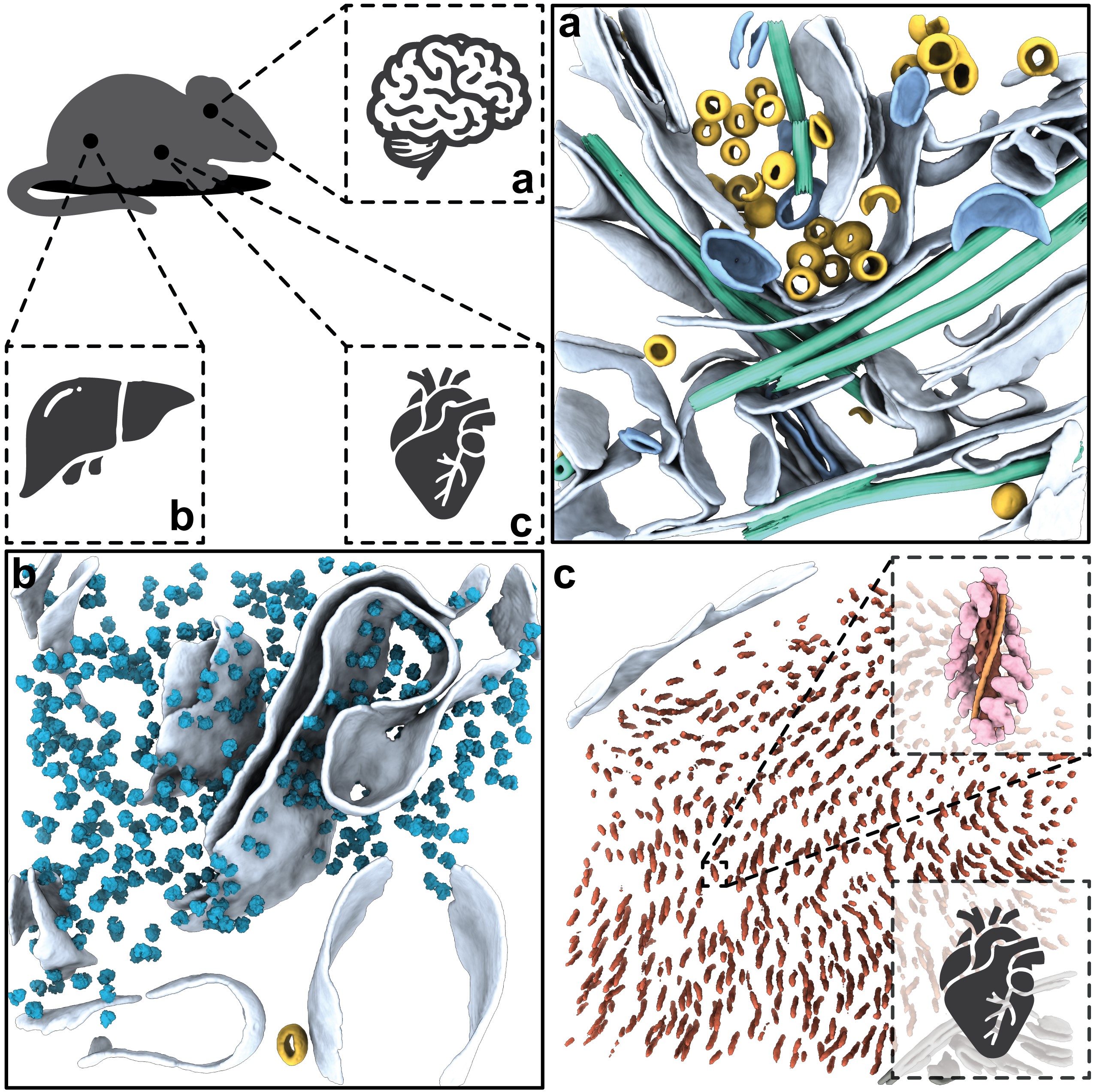
To determine the scope of their new approach, the Human Technopole researchers tested SOLIST on various biological samples like human brain organoids, mouse liver, brain, and heart tissues allowing them to see the natural organization of e.g. nerves and muscle fibers (watch the video 2). They found that SOLIST was more efficient and approachable than previous methods, allowing even novice users to generate better data in less time.
The scientists said their new approach gave them but a first glimpse at developing human brain organoids, an important model system at HT to study human central nervous system disorders such as Alzheimer’s and Parkinson’s. While it is not yet as efficient as mechanical sectioning at room temperature, SOLIST is a significant improvement over previous cryo-techniques, which had low throughput and suffered from ice contamination and poor sample stability.
The study leader, Philipp Erdmann, and his team are convinced that SOLIST will soon help create high-resolution datasets for more complex samples like patient tissues. This will advance our understanding of healthy and disease-related molecular processes in humans, moving us closer to a “biopsy at the nanoscale.”
Nguyen, H.T.D., Perone, G., Klena, N. et al. Serialized on-grid lift-in sectioning for tomography (SOLIST) enables a biopsy at the nanoscale. Nat Methods 21, 1693–1701 (2024). https://doi.org/10.1038/s41592-024-02384-6
Share:
You could also like:
-

FRRB funds the Glastonbury Group to study the immune landscape of pancreatic cancer
The Glastonbury Group at Human Technopole has been awarded a competitive grant as part of the “From Bed To Bench: the way to innovation” funding programme promoted by the Fondazione Regionale per la Ricerca Biomedica (FRRB). The study, funded with 2.000.000 euros, 600.000 of which will go to the Glastonbury group directly, will begin in late 2025 and run for 3 years.
-
Automating the science of how microbes grow
Human Technopole researchers have developed Kinbiont, an open-source software that automates the analysis of microbial growth data and returns quantitative, interpretable models of microbial behaviour. The results are published in Nature Communications.
-
How the human genome condenses during cell division
As a result of an international collaboration, Human Technopole researchers identify M18BP1 as an activator of condensin II through a phosphorylation-regulated competition with MCPH1. This previously unknown mechanism fills a significant gap in our understanding of cell division and suggests avenues for exploring how its misregulation contributes to human disease. The results of the research are published in Molecular Cell.
-
Ageing and Chronic Diseases: Europe Rewards Human Technopole’s Research
The ERC has awarded HT a €2.5 million grant for a study investigating how lifestyle, biology and environmental exposures weaken the immune system over the course of life and increase vulnerability to disease in old age. This new knowledge will help develop models to predict long-term disease risk.
-

1 fully-funded PhD Position in the Jug Group through DADS
Are you passionate about AI, cutting-edge research, and making a real impact in biomedical science? The Jug Group at Human Technopole is thrilled to announce a new PhD opportunity as part of the prestigious PhD Programme in Data Analytics and Decision Sciences.