National Facility for Structural Biology
Every biological process, including physiological and pathological events, is precisely orchestrated by active and reactive biological macromolecules. The function, organization, and activity of these molecules strictly depend on both their three-dimensional structure and the cellular environment in which they operate. In this context, the National Facility for Structural Biology stands as a one-of-a-kind scientific and technological hub for integrative structural biology.
The NF for Structural Biology consists of six Infrastructural Units (IU) working together to (i) provide and support conventional practices, and (ii) promote and establish innovative workflows in the field of integrative structural biology. Specifically, the Facility foresees the following Units:
-
- IU1 – Cryo-Electron Microscopy: This Unit aims at identifying, visualizing, and characterizing biological players of interest, both isolated and within their cellular compartments.
- IU2 – Biomass Production: This Unit provides access to different cell lines for protein expression and performs scale-up of bioprocesses for large-scale productions.
- IU3 – Biophysics: This Unit is a technological platform for biophysical characterization of macromolecules and their interactions
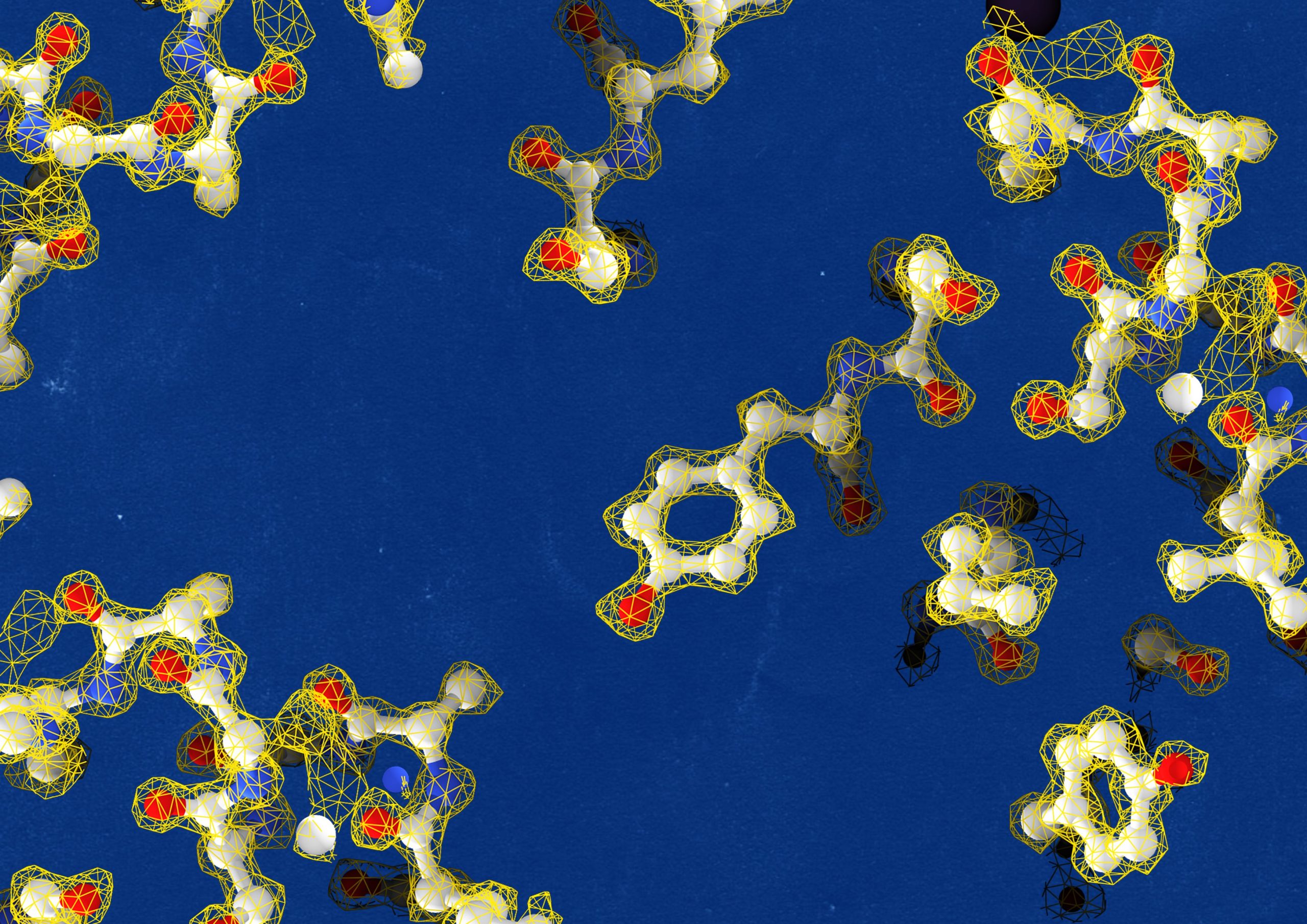
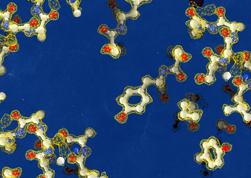
- IU4 – Structural Proteomics: This Unit relies on crosslinking mass spectrometry (XL-MS) to provide topological and structural restraints on protein-protein interactions in samples ranging from purified protein complexes to cellular fractions.
- IU5 – Dynamic Single-molecule: This Unit provides tool to visualise biological processes in real-time with single-molecule sensitivity thanks to cutting-edge instruments that combine optical tweezers with fluorescence and label-free detection modules.
- IU6 – Technology Development: This Unit, planned to be operational in 2026-2028, will be where all other units converge when it comes to pushing technological limitations in the field of integrative structural biology.
The mission of the National Facility for Structural Biology is to provide access to a highly productive, worldclass scientific hub capable of addressing disease mechanisms across scales, from tissue to amino acid side-chains, in contemporary life science.
Open Call
25-SB-ROUND2 - National Facility for Structural Biology
The call for Access for the National Facility for Structural Biology (Call ID: 25-SB-ROUND2) is open from 1 June to 30 September 2025.
Details:
For general enquiries about the call: [email protected]
For technical enquiries about the services: [email protected]
Downloads:
Links:
Services
- Negative Stain EM Screening
- Cryo-EM Screening
- High-resolution Cryo-TEM Imaging
- Volume Electron Microscopy
- Cryo-FM Imaging
- Protein expression in insect cells
- Protein expression in mammalian cells in suspension
- Protein Expression in yeast and bacteria cells
- Characterisation of Macromolecular Samples
- Measurement of affinity constants
- Sample Check/Optimisation for Structural Biology Workflows
- Protein purification
- Crosslinking MS acquisition of purified protein complex without crosslinking reaction optimisation
- Crosslinking MS acquisition of purified protein complex with crosslinking reaction optimisation
- Crosslinking MS acquisition of immunoprecipitate from tagged overexpressed bait
- Crosslinking MS acquisition of immunoprecipitate from endogenous material or of cellular fraction
- Integrative modeling with crosslinking MS and cryo-EM data
- Proteomics acquisition on Orbitrap Astral – high load
- Proteomics acquisition on Orbitrap Astral – high throughput
- Assay Development
- Data Acquisition
Instruments
UI1 – Cryo-Electron Microscopy Unit
![]()
IU2 – Biomass Production Unit
![]()
IU3 – Biophysics Unit
![]()
IU4 – Structural Proteomics Unit
![]()
IU5 – Dynamic Single Molecule Unit
![]()
Facility Members
-
 Paolo Swuec
Head of National Facility for Structural Biology -
 Joanna Jadwiga Andrecka
Senior Manager - Single Molecule Support Unit -
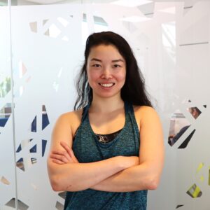 Si Hui Elisa Chen
Electron Microscopy Specialist -
 Nataliya Danilenko
Technician, Biophysics Scientific Service Unit -
 Alessio Di Ianni
Technician -
 Gaetano D’Urso
Electron Microscopy Specialist -
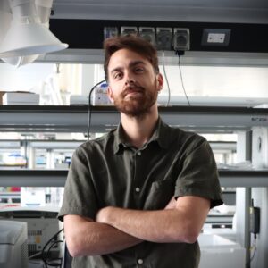
 Andrea Graziadei
Senior Manager - Structural Proteomics -
 Gabriele Marcassa
Technician -
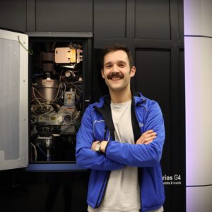
 Marko Nedeljkovic
Senior Technician -
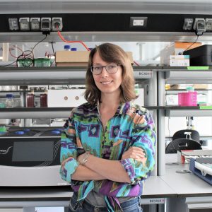
 Sebastiano Pasqualato
Senior Manager Biophysics -
 Pedro Miguel Pereira Machado
Electron Microscopy Specialist -
 Alessandro Scardua
Senior Manager Biomass Production & Kitchen -
 Simona Sorrentino
Electron Microscopy Specialist -
 Janine Weber
Senior Technician Biomass Production