A new model system for cortical development in vitro
Researchers from Human Technopole, the Institute of Molecular Biotechnology and Bicocca University established a method for developing brain assembloids that allows reproducing salient aspects of the antero-posterior polarity of the human cerebral cortex in vitro and opens new possibilities for disease modelling. The study is published in Nature Methods.
The cerebral cortex – the outermost layer of the brain – plays critical roles in memory, thinking, learning, reasoning, and sense. Its development, or corticogenesis, requires the concerted expression of transcription factors and the activation of signalling cascades for different areas and functions to mature properly.
Human Pluripotent Stem Cell (hPSC)-derived brain organoids have been extensively used to study corticogenesis and disease-related alterations. However, these models lack the typical anterior-to-posterior organisation and diversity of cell types observed in vivo.
To address this issue, the groups of Giuseppe Testa – Head of the Neurogenomics at Human Technopole and professor at the University of Milan – Veronica Krenn – Human Technopole Early Career Fellow and Group Leader at University of Milan-Bicocca – and Jürgen Knoblich – Deputy Scientific Director of the Institute of Molecular Biotechnology (IMBA) of the Austrian Academy of Sciences – joined forces to developed a new protocol for engineering cortical organoids that recapitulate salient aspects of the polarized transcriptional and signalling patterns observed in vivo.
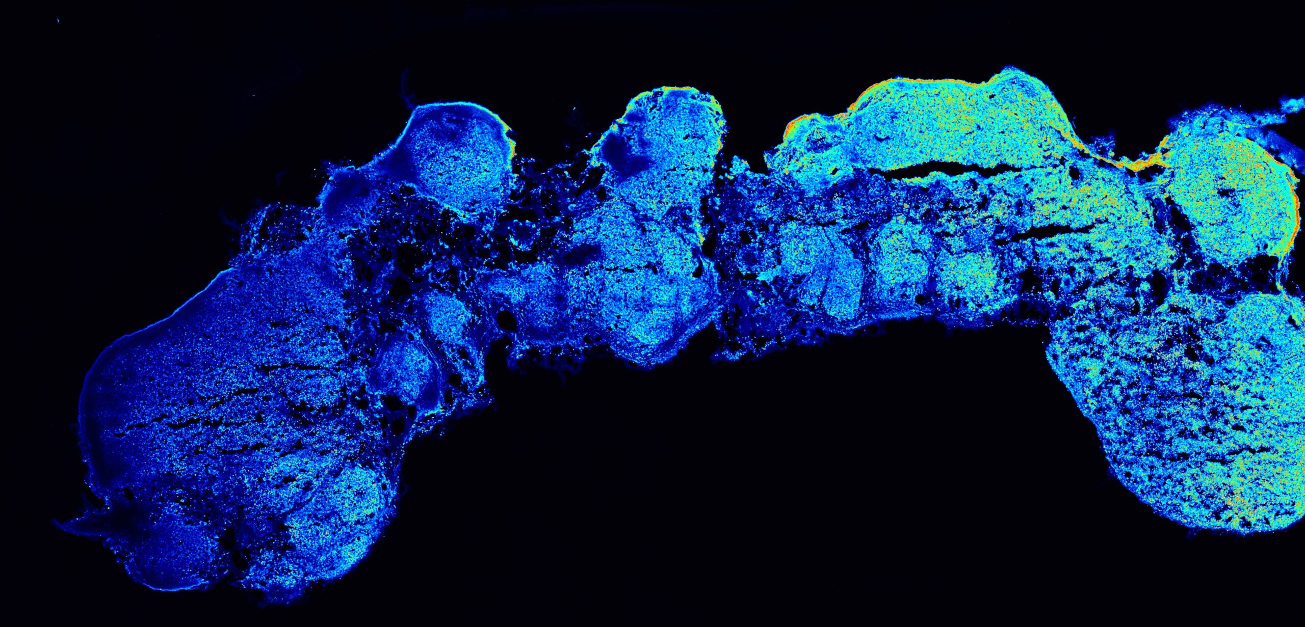
Leveraging the observation that fibroblast growth factor 8 (FGF8) induces a dose-dependent expression of anterior and posterior markers in round cortical organoids, the researchers generated mosaic bodies (mosaic organizer-like embryoid bodies, OrEBs or assembloids) consisting of hPSC-derived brain organoids fused with hPSCs that constitutively produced and secreted FGF8. Such assembloids showed the features of anterior telencephalic development. Based on these findings, the teams increased the length of the organoids in the mosaic bodies to boost the gradient of FGF8 and enable position-dependent effects of FGF8 expression. Strikingly, they observed significant differences in the expression of anterior and posterior markers and fate acquisition at opposite poles of the assembloids. RNA-sequencing analysis on dissected proximal (P), medial (M) and distal (D) segments of individual assembloids confirmed that the P-to-D FGF8 gradient partly induced the in vivo antero-posterior gene expression pattern. Like the developing human brain, each segment showed a different cell-type composition as revealed by single-cell RNA-sequencing. Finally, the researchers focused on Fibroblast growth factor receptor 3 (FGFR3), one of the hits that showed low-to-high P-to-D expression in the assembloids. Mutations in FGFR3 are known to cause achondroplasia, a genetic disorder having as a primary feature dwarfism. Achondroplasia-related mutation in FGFR3 (G380R) decreased the position-dependent proliferation effects along the P-to-D axis of the assembloids.
“Unlike previous attempts that relied on poorly controlled and low-frequency sources of morphogens resulting in local patterning effects in individual organoids, our approach utilises a well-controlled source of FGF8 and culture media formulations with minimal exogenous signals to consistently generate cortical polarity along an organoid’s entire longitudinal axis”, the authors say.
Giuseppe Testa, one of the leaders of this research, also commented, “Polaraised cortical assembloids capture in a dish what had been predicted by the “protomap” model, namely that the diversity of cells from different regions of our cortex is primed early on in development. We are excited about the transformative opportunity that this method now affords to study how genetic and environmental factors contribute to neuropsychiatric disorders by acting on such critical early events”.
In the image: a polarized cortical assembly showing a spatial gradient of expression along its longitudinal axis (from left to right, colors from blue to yellow-red). @Camilla Bosone (IMBA)
Share:
You could also like:
-
HT National Facilities: One Year On, New Call Open
On 10 June, Human Technopole marks the one-year anniversary of the official opening of its National Facilities, a key milestone in our mission to support excellence in life sciences across Italy. Celebrating this important date, we are pleased to announce that the new Call for Access to the National Facilities is now open. Researchers can submit their proposals until 30 September 2025.
-
10 Fully Funded PhD Scholarships in Systems Medicine through SEMM
Human Technopole is offering up to 10 fully funded PhD scholarships through the SEMM PhD Program in Systems Medicine. These scholarships are open to talented and motivated graduates—both from Italy and abroad – interested in pursuing doctoral research.
-

Human Technopole Commitment to Gender Equality
Human Technopole is proud to announce the launch of its new Gender Equality Plan (GEP) for 2025-2027, a strategic initiative designed to further integrate gender equality across all aspects of the Institute, building upon the successful outcomes of the previous GEP (2022-2024). HT has also achieved the UNI/PdR 125:2022 certification, which recognizes organizations that promote gender equality and foster inclusive workplaces.
-

2 fully funded PhD fellowships at HT through SISSA
Human Technopole is offering 2 fully funded 4-year PhD fellowships to young scientists from the national and international community who wish to undertake a doctoral degree on a project focused on Computational Biology.
-

Childhood Cancer: Free DNA in Blood Reveals Therapy Resistance
An international study coordinated by Milan’s Human Technopole, the Institute of Cancer Research (ICR) in London, and the Royal Marsden Hospital in London has shown that certain DNA fragments found in the blood of paediatric cancer patients can be used as “biomarkers” to obtain information on the characteristics of the disease and its ability to resist therapies. Analysing these fragments could represent an effective alternative to tumour tissue biopsy, a practice that is particularly difficult in children.