National Facility for Light Imaging
The National Facility for Light Imaging offers services related to imaging, sample preparation, functional imaging and cell sorting. The mission of the facility is to support science: to reach this goal, the facility and its team are available to internal and external users, providing support for designing experiments, acquiring data on the facility’s cutting-edge scientific instruments, offering training and development of new protocols and technologies. Integrated pipelines with other facilities of Human Technopole allow users to access streamlined multi-facility workflows, where data acquired in the National Facility for Light Imaging can be further processed and analyzed by other National Facilities.
Currently, four infrastructural Units (IU) within the National Facility are active and available to external applicants:
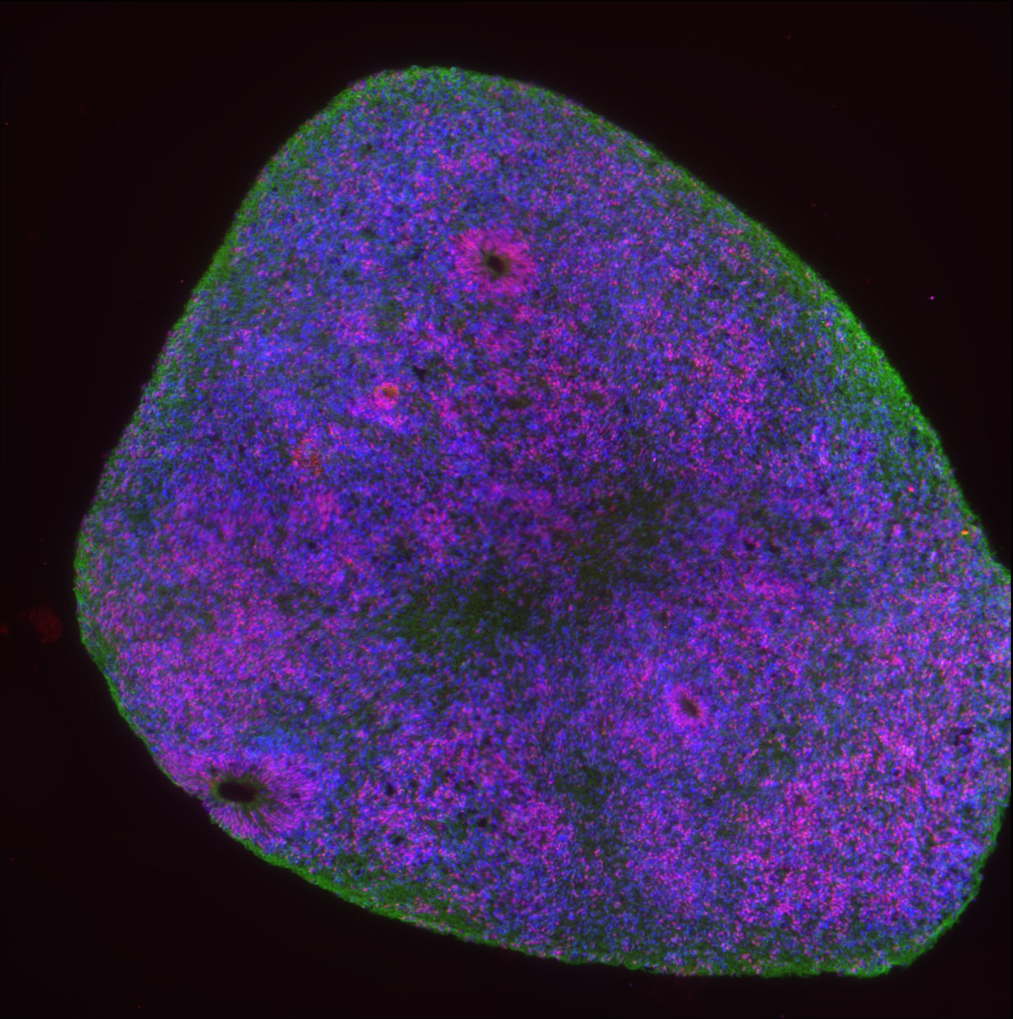
- IU1 – Imaging: The Imaging IU offers access to several high-end microscopy systems. The microscopes can be used to image fixed or living samples across spatial scales, ranging from super-resolution sub-cellular imaging to whole organ imaging. After a mandatory training, users can access the microscopes autonomously. Supervision and assistance from the unit’s staff is available upon request.
- IU 2 – Tissue Processing: The Tissue Processing IU provides platforms, technical support and training for preparing samples for downstream analysis, such as light imaging or spatial transcriptomics.
- IU 3 – Flow cytometry applications: The Flow Cytometry IU offers full-service sorting & cloning of rare cellular populations, particle enrichment, and high purity bulk sorts. The unit also offers Analysis Services including combined scalar and image capabilities, and advice in experimental design and training.
- IU 4 – High-content imaging: Service under construction
- IU 5 – Ion Imaging: The Ion Imaging IU offers functional cell imaging services using fluorescence-based time-lapse recordings of intracellular ion oscillations or optics-free extracellular voltage recordings through microelectrode arrays (MEA) systems. Optical imaging is primarily based on confocal or epifluorescence microscopy
- IU 6 – Technology Development – custom microscopy: Service under construction

Open Call
25-LI-ROUND2 - National Facility for Light Imaging
The call for Access for the National Facility for Light Imaging (Call ID: 25-LI-ROUND2) is open from 1 June to 30 September 2025.
Details:
For general enquiries about the call: [email protected]
For technical enquiries about the services: [email protected]
Downloads:
Links:
Services
Wide-field
- Leica Thunder Imager Live Cell
- Zeiss Axioscope5
- Zeiss Axiozoom V16 with apotome
Confocal
- Zeiss LSM980
- Zeiss LSM980-NLO
- Nikon Ti2 with CREST v3 spinning disk scan head
- Leica Stellaris 8 for live-cell imaging
Super-resolution
- Zeiss Elyra7
- Abberrior FacilityLine
- Leica Stellaris 8 for super-resolution imaging
Lightsheet
- Zeiss Lattice Lightsheet 7
Image Analysis can be provided as a combined service by the National Facility for Data Handling and Analysis.
Slide Scanner
- Zeiss Axioscan Z.1 automated slide scanner
Image Analysis can be provided as a combined service by the National Facility for Data Handling and Analysis.
Flow Cytometry Cell Sorting
- BD FACSDiscover S8
- MoFlo Astrios EQ
Flow Cytometry Analysis
- Cytek Aurora Spectral Analyzer
- ImageStream Mark II Imaging Flow Cytometer
- CytoFLEX LX Flow Cytometer
Assisted Flow Cytometry Analysis
Consultation session: Sample Preparation/Panel Design/Data Analysis
Ion imaging assisted experiment
- MEA assay
- Ion Imaging assay
Instruments
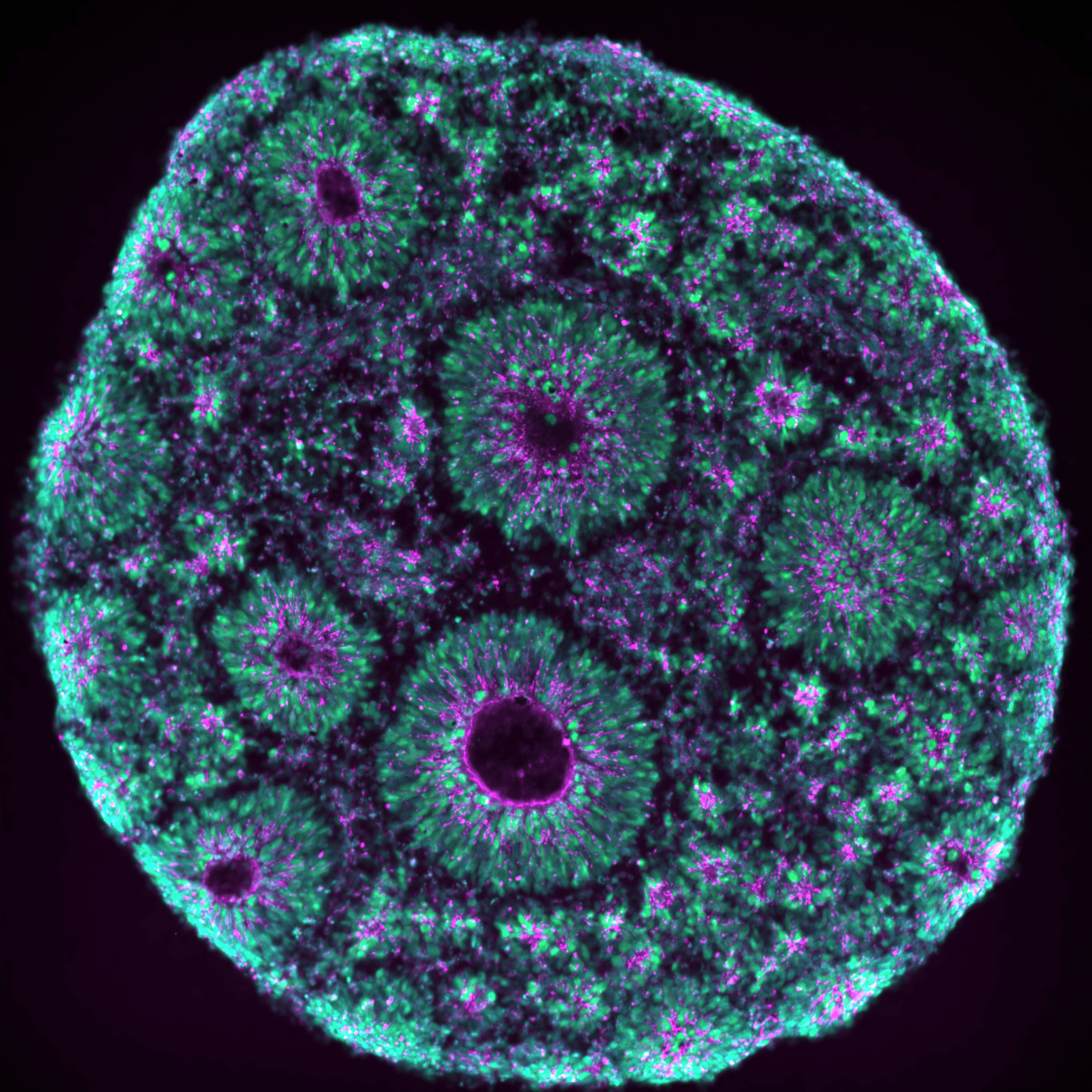
IU1 Imaging
![]()
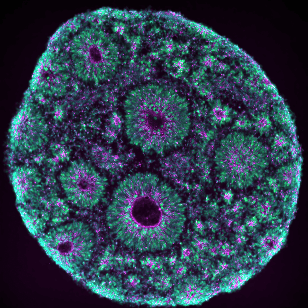
IU2 Tissue Processing
![]()
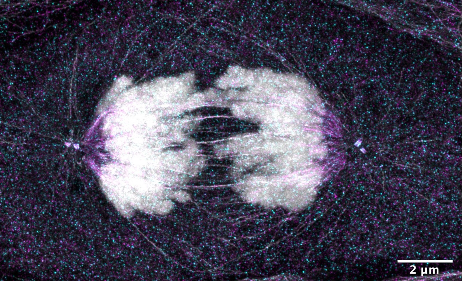
IU3 Flow Cytometry
![]()
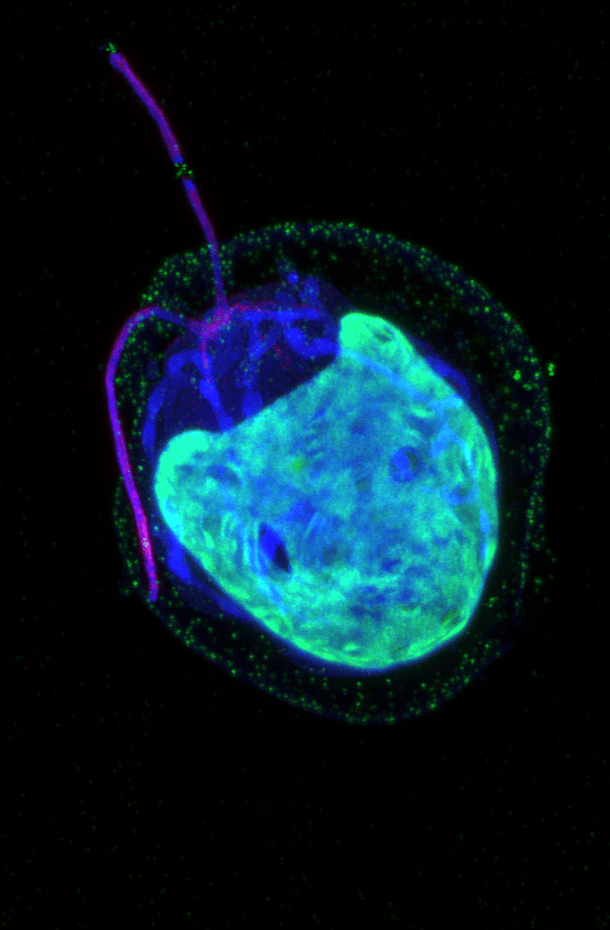
IU5 Ion Imaging
![]()
Facility Members
-
 Nicola Maghelli
Head of National Facility for Light Imaging -
 Erica Belgio
Senior Technician -
 Silvia Bombelli
Senior Technician -
 Emanuele Canonico
Senior Technician -
 Francesca Casagrande
Senior Technician Light Imaging Facility -
 Alessandro Di Maio
Senior Technician -
 Alessandra Fasciani
Senior Technician -
 Ilaria Laface
Technician -
 Filippo Mirabella
Senior Technician -
 Alessio Palini
Senior Manager Flow Cytometry Core Facility -
 Nicolò Panini
Senior Technician -
 Diletta Pozzi
Manager – Electrophysiology Scientific Service -
 Dario Ricca
Senior Technician