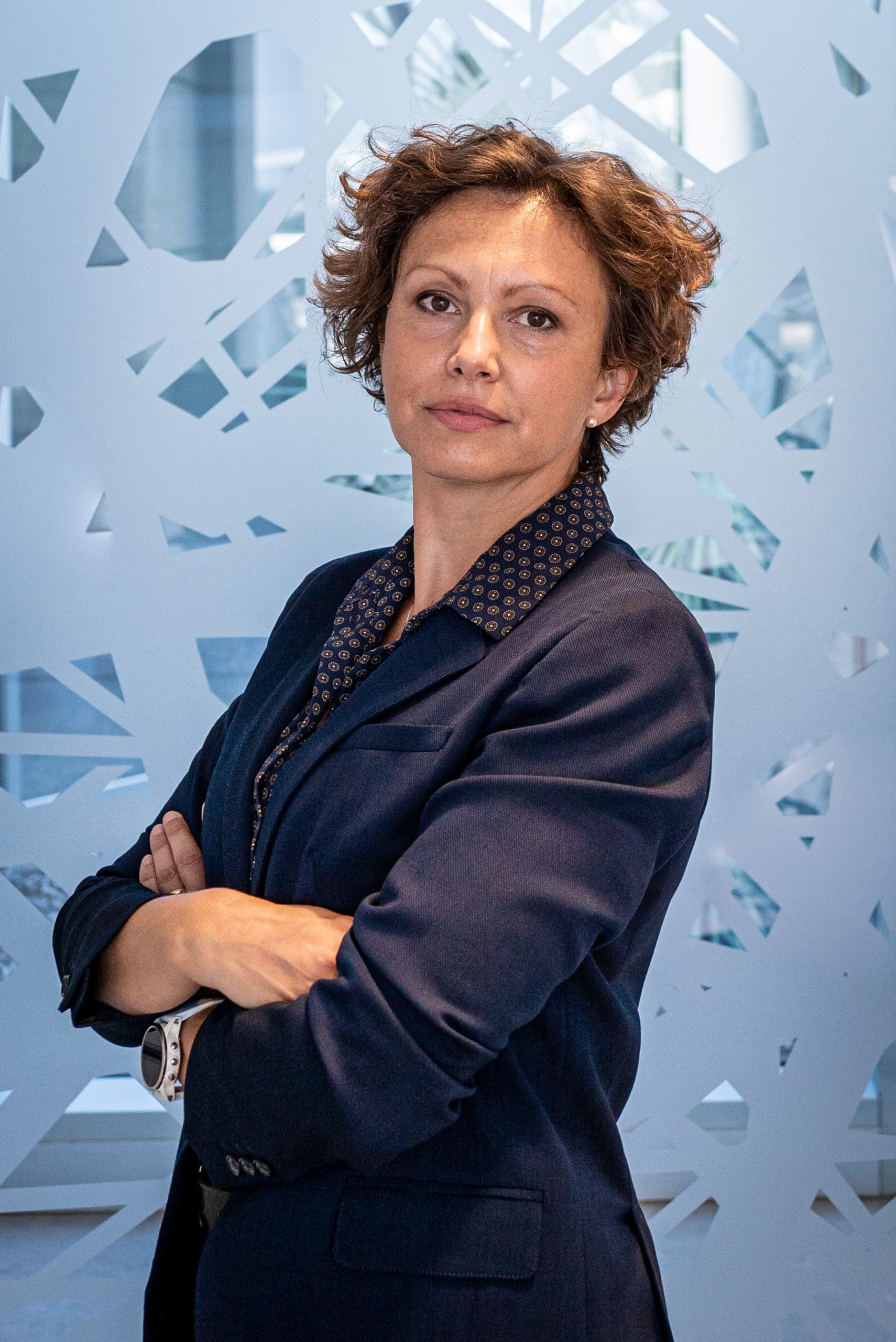
Gaia Pigino
- Associate Head of Structural Biology Research Centre, Structural biology
- Research Group Leader, Pigino Group
Gaia Pigino is a biologist, currently Associate Head of the Structural Biology Center at Human Technopole, after 9 years as Research Group Leader at the Max Planck Institute CBG in Dresden. She collaborate with Alessandro Vannini to develop the Centre for Structural Biology. Gaia’s laboratory studies molecular mechanisms and principles of self-organisation in cilia and other subcellular structures that are of fundamental importance for human health and disease.
CURRENT POSITION
| Since 2021 | Associate Head of the Structural Biology Center at Human Technopole, Milan, Italy |
| Since 2012 | Research Group Leader at MPI-CBG, the Max Planck Institute of Molecular Cell Biology and Genetics, Dresden, Germany |
POSTDOCTORAL RESEARCH
| 2010-2012 | Postdoctoral EMBO Long Term fellow Laboratory of Biomolecular Research (BMR), Department of Biology and Chemistry, Paul Scherer Institute (PSI) Switzerland. Supervisor: Prof. T. Ishikawa. |
| 2009-2011 | Postdoctoral researcher Institute for Molecular Biology and Biophysics, Swiss Federal Institute of Technology (ETH Zürich), Zurich, Switzerland. Supervisor: Prof. T. Ishikawa. |
| 2007-2009 | Postdoctoral MIUR research fellow Fellowship of the “Ministero Italiano dell’Istruzione, dell’Università e della Ricerca”. Laboratory of Cryotechniques for Electron Microscopy, Department of Evolutionary Biology, University of Siena. Supervisor: Prof. P. Lupetti. |
| 2009 | Participant at the Physiology Course at MBL in Woods Hole Marine Biological Laboratory, Woods Hole. Directors: Dyche Mullins and Claire Waterman. |
EDUCATION
| 2003-2007 | Ph.D. Student (Ph.D. Fellowship by the Italian government “Ministero Italiano dell’Istruzione, dell’Università e della Ricerca”). Department of Evolutionary Biology, University of Siena. Supervisor: Prof. F. Bernini and Prof. C. Leonzio. |
| 2002 | Diploma in Natural Science (Summa cum laude). University of Siena, Italy. Thesis supervisors: Prof. C. Leonzio and Prof. F. Bernini. |
OTHER POSITIONS
| 2003 | Research Associate. Department of Environmental Sciences G. Sarfatti, University of Siena. Advisor: Prof. C. Leonzio |
AWARDS and FUNDING
| 2022 | EMBO Member |
| 2019 | DFG Grant – GAČR-DFG Cooperation |
| 2018 | PoL starting fellowship (from the Dresden Excellence Cluster ‘Physics of Life’) |
| 2018 | Keith R. Porter Fellow Award for Cell Biology |
| 2018 | ERC Consolidator Grant (ERC-2018-COG N#819826 CiliaTubulinCode) |
| 2018 | Excellence Cluster ‘Physics of Life’, as a core Principal Investigator |
| 2010 | EMBO Long Term fellowship |
| 2009 | Scholarship from the Marine Biological Laboratory (Woods Hole, Massachusetts) MBL Physiology Course. |
| 2007 | Post-Doctoral Research fellowship from MIUR. |
| 2003 | Ph.D. Fellowship from MIUR. |
Fellowship to students and postdocs
| 2022 | EMBO Long Term Fellowship to Helen Foster |
| 2021 | EMBO Postdoc Fellowship to Nikolai Klena |
| 2019 | HFSP Postdoc Fellowship to Adrian Nievergelt |
| 2018 | EMBO Long Term Fellowship to Adrian Nievergelt |
| 2017 | Marie Curie Fellowship to Adam Schröfel (H2020-MSCA-IF-2016) |
| 2015 | DIGS-BB Fellowship to Guendalina Marini |
| 2012 | DIGS-BB Fellowship to Ludek Stepanek |
Google Scholar
Contacts
Follow on
Publications
-
11/2021 - eLife
A WDR35-dependent coat protein complex transports ciliary membrane cargo vesicles to cilia
Intraflagellar transport (IFT) is a highly conserved mechanism for motor-driven transport of cargo within cilia, but how this cargo is selectively transported to cilia and across the diffusion barrier is unclear. WDR35/IFT121 is a component of the IFT-A complex best known for its role in ciliary retrograde transport. In the absence of WDR35, small mutant […]
-
08/2021 - Journal of Cell Science
In vivo imaging shows continued association of several IFT A, B and dynein complexes while IFT trains U-turn at the tip
Flagellar assembly depends on intraflagellar transport (IFT), a bidirectional motility of protein carriers, the IFT trains. The trains are periodic assemblies of IFT-A and IFT-B subcomplexes and the motors kinesin-2 and IFT dynein. At the tip, anterograde trains are remodeled for retrograde IFT, a process that in Chlamydomonas involves kinesin-2 release and train fragmentation. However, […]
-
06/2021 - Journal of Cell Science
The structural basis of intraflagellar transport at a glance
The intraflagellar transport (IFT) system is a remarkable molecular machine used by cells to assemble and maintain the cilium, a long organelle extending from eukaryotic cells that gives rise to motility, sensing and signaling. IFT plays a critical role in building the cilium by shuttling structural components and signaling receptors between the ciliary base and […]
-
05/2021 - Current Biology
Intraflagellar transport
Cells need to be able to sense different types of signals, such as chemical and mechanical stimuli, from the extracellular environment in order to properly function. Most eukaryotic cells sense these signals in part through a specialized hair-like organelle, the cilium, that extends from the cell body as a sort of antenna. The signaling and […]
-
03/2021 - BioRxiv
Intraflagellar transport trains can turn around without the ciliary tip complex
Cilia and flagella are microtubule doublet based organelles found across the eukaryotic tree of life. Their very high aspect ratio and crowded interior are unfavourable to diffusive transport for their assembly and maintenance. Instead, a highly dynamic system of intraflagellar transport (IFT) trains moves rapidly up and down the cilium. However, the mechanism of how […]