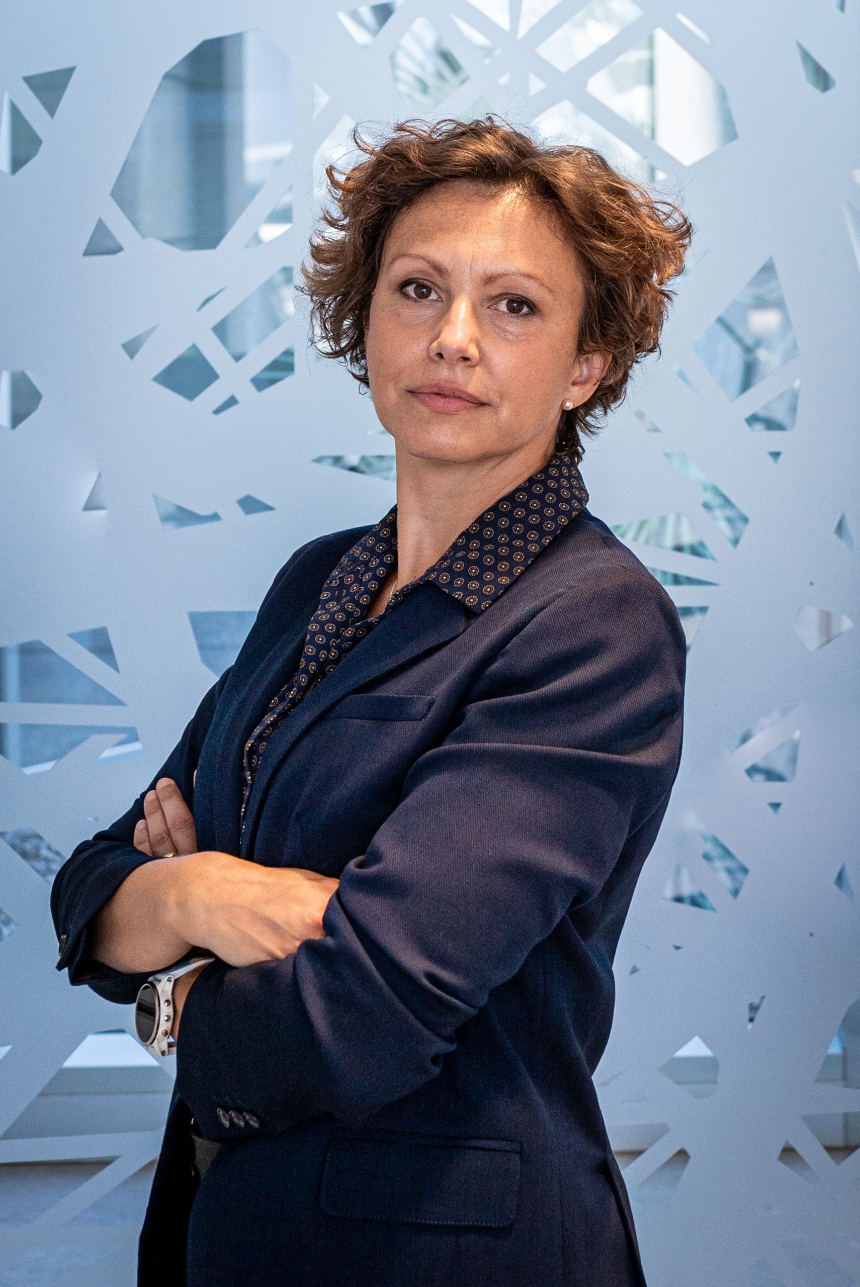
Gaia Pigino
- Associate Head of Structural Biology Research Centre, Structural biology
- Research Group Leader, Pigino Group
Gaia Pigino is a biologist, currently Associate Head of the Structural Biology Center at Human Technopole, after 9 years as Research Group Leader at the Max Planck Institute CBG in Dresden. She collaborate with Alessandro Vannini to develop the Centre for Structural Biology. Gaia’s laboratory studies molecular mechanisms and principles of self-organisation in cilia and other subcellular structures that are of fundamental importance for human health and disease.
CURRENT POSITION
| Since 2021 | Associate Head of the Structural Biology Center at Human Technopole, Milan, Italy |
| Since 2012 | Research Group Leader at MPI-CBG, the Max Planck Institute of Molecular Cell Biology and Genetics, Dresden, Germany |
POSTDOCTORAL RESEARCH
| 2010-2012 | Postdoctoral EMBO Long Term fellow Laboratory of Biomolecular Research (BMR), Department of Biology and Chemistry, Paul Scherer Institute (PSI) Switzerland. Supervisor: Prof. T. Ishikawa. |
| 2009-2011 | Postdoctoral researcher Institute for Molecular Biology and Biophysics, Swiss Federal Institute of Technology (ETH Zürich), Zurich, Switzerland. Supervisor: Prof. T. Ishikawa. |
| 2007-2009 | Postdoctoral MIUR research fellow Fellowship of the “Ministero Italiano dell’Istruzione, dell’Università e della Ricerca”. Laboratory of Cryotechniques for Electron Microscopy, Department of Evolutionary Biology, University of Siena. Supervisor: Prof. P. Lupetti. |
| 2009 | Participant at the Physiology Course at MBL in Woods Hole Marine Biological Laboratory, Woods Hole. Directors: Dyche Mullins and Claire Waterman. |
EDUCATION
| 2003-2007 | Ph.D. Student (Ph.D. Fellowship by the Italian government “Ministero Italiano dell’Istruzione, dell’Università e della Ricerca”). Department of Evolutionary Biology, University of Siena. Supervisor: Prof. F. Bernini and Prof. C. Leonzio. |
| 2002 | Diploma in Natural Science (Summa cum laude). University of Siena, Italy. Thesis supervisors: Prof. C. Leonzio and Prof. F. Bernini. |
OTHER POSITIONS
| 2003 | Research Associate. Department of Environmental Sciences G. Sarfatti, University of Siena. Advisor: Prof. C. Leonzio |
AWARDS and FUNDING
| 2022 | EMBO Member |
| 2019 | DFG Grant – GAČR-DFG Cooperation |
| 2018 | PoL starting fellowship (from the Dresden Excellence Cluster ‘Physics of Life’) |
| 2018 | Keith R. Porter Fellow Award for Cell Biology |
| 2018 | ERC Consolidator Grant (ERC-2018-COG N#819826 CiliaTubulinCode) |
| 2018 | Excellence Cluster ‘Physics of Life’, as a core Principal Investigator |
| 2010 | EMBO Long Term fellowship |
| 2009 | Scholarship from the Marine Biological Laboratory (Woods Hole, Massachusetts) MBL Physiology Course. |
| 2007 | Post-Doctoral Research fellowship from MIUR. |
| 2003 | Ph.D. Fellowship from MIUR. |
Fellowship to students and postdocs
| 2022 | EMBO Long Term Fellowship to Helen Foster |
| 2021 | EMBO Postdoc Fellowship to Nikolai Klena |
| 2019 | HFSP Postdoc Fellowship to Adrian Nievergelt |
| 2018 | EMBO Long Term Fellowship to Adrian Nievergelt |
| 2017 | Marie Curie Fellowship to Adam Schröfel (H2020-MSCA-IF-2016) |
| 2015 | DIGS-BB Fellowship to Guendalina Marini |
| 2012 | DIGS-BB Fellowship to Ludek Stepanek |
Google Scholar
Contacts
Follow on
Publications
-
07/2019 - Current Opinion in Structural Biology
Towards a mechanistic understanding of cellular processes by cryoEM
A series of recent hardware and software developments have transformed cryo-electron microscopy (cryoEM) from a niche tool into a method that has become indispensable in structural and functional biology. Samples that are rapidly frozen are encased in a near-native state inside a layer of amorphous ice, and then imaged in an electron microscope cooled to […]
-
07/2019 - Methods in Cell Biology
Content-aware image restoration for electron microscopy
Multiple approaches to use deep neural networks for image restoration have recently been proposed. Training such networks requires well registered pairs of high and low-quality images. While this is easily achievable for many imaging modalities, e.g., fluorescence light microscopy, for others it is not. Here we summarize on a number of recent developments in the […]
-
06/2019 - Methods in Cell Biology
Yeast membraneless compartments revealed by correlative light microscopy and electron tomography
Yeast essential enzymes are able to assemble and form membrane-less compartments in the cytoplasm during stress conditions (Narayanaswamy et al., 2009). These microcompartments form rapidly under ATP-depletion upon cellular regulation of pH and molecular crowding (Munder et al., 2016). So far, the behavior of most of these enzymes has been characterized by live imaging using fluorescence […]
-
05/2019 - Methods in Cell Biology
In situ cryo-electron tomography and subtomogram averaging of intraflagellar transport trains
In situ cryo-electron tomography (cryo-ET) and subtomogram averaging are powerful tools, able to provide 3D structures of biological samples at sub-nanometer resolution, while preserving information about cellular context and higher-order assembly. Best results are typically achieved, when applied to highly repetitive structures, such as viruses. Other typical examples are protein complexes that decorate long stretches along ciliary microtubules at […]
-
11/2018 - Nature Cell Biology
The cryo-EM structure of intraflagellar transport trains reveals how dynein is inactivated to ensure unidirectional anterograde movement in cilia
Movement of cargos along microtubules plays key roles in diverse cellular processes, from signalling to mitosis. In cilia, rapid movement of ciliary components along the microtubules to and from the assembly site is essential for the assembly and disassembly of the structure itself1. This bidirectional transport, known as intraflagellar transport (IFT)2, is driven by the […]