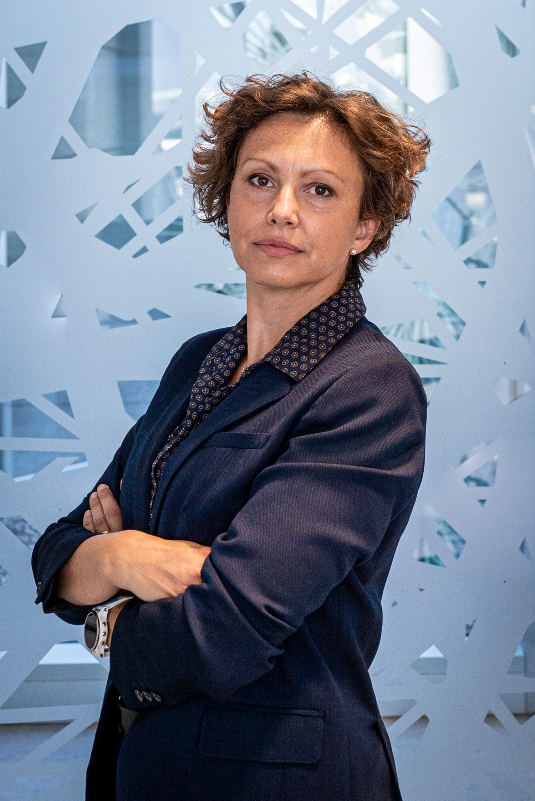
Gaia Pigino
- Associate Head of Structural Biology Research Centre, Structural biology
- Research Group Leader, Pigino Group
Gaia Pigino is a biologist, currently Associate Head of the Structural Biology Center at Human Technopole, after 9 years as Research Group Leader at the Max Planck Institute CBG in Dresden. She collaborate with Alessandro Vannini to develop the Centre for Structural Biology. Gaia’s laboratory studies molecular mechanisms and principles of self-organisation in cilia and other subcellular structures that are of fundamental importance for human health and disease.
CURRENT POSITION
| Since 2021 | Associate Head of the Structural Biology Center at Human Technopole, Milan, Italy |
| Since 2012 | Research Group Leader at MPI-CBG, the Max Planck Institute of Molecular Cell Biology and Genetics, Dresden, Germany |
POSTDOCTORAL RESEARCH
| 2010-2012 | Postdoctoral EMBO Long Term fellow Laboratory of Biomolecular Research (BMR), Department of Biology and Chemistry, Paul Scherer Institute (PSI) Switzerland. Supervisor: Prof. T. Ishikawa. |
| 2009-2011 | Postdoctoral researcher Institute for Molecular Biology and Biophysics, Swiss Federal Institute of Technology (ETH Zürich), Zurich, Switzerland. Supervisor: Prof. T. Ishikawa. |
| 2007-2009 | Postdoctoral MIUR research fellow Fellowship of the “Ministero Italiano dell’Istruzione, dell’Università e della Ricerca”. Laboratory of Cryotechniques for Electron Microscopy, Department of Evolutionary Biology, University of Siena. Supervisor: Prof. P. Lupetti. |
| 2009 | Participant at the Physiology Course at MBL in Woods Hole Marine Biological Laboratory, Woods Hole. Directors: Dyche Mullins and Claire Waterman. |
EDUCATION
| 2003-2007 | Ph.D. Student (Ph.D. Fellowship by the Italian government “Ministero Italiano dell’Istruzione, dell’Università e della Ricerca”). Department of Evolutionary Biology, University of Siena. Supervisor: Prof. F. Bernini and Prof. C. Leonzio. |
| 2002 | Diploma in Natural Science (Summa cum laude). University of Siena, Italy. Thesis supervisors: Prof. C. Leonzio and Prof. F. Bernini. |
OTHER POSITIONS
| 2003 | Research Associate. Department of Environmental Sciences G. Sarfatti, University of Siena. Advisor: Prof. C. Leonzio |
AWARDS and FUNDING
| 2022 | EMBO Member |
| 2019 | DFG Grant – GAČR-DFG Cooperation |
| 2018 | PoL starting fellowship (from the Dresden Excellence Cluster ‘Physics of Life’) |
| 2018 | Keith R. Porter Fellow Award for Cell Biology |
| 2018 | ERC Consolidator Grant (ERC-2018-COG N#819826 CiliaTubulinCode) |
| 2018 | Excellence Cluster ‘Physics of Life’, as a core Principal Investigator |
| 2010 | EMBO Long Term fellowship |
| 2009 | Scholarship from the Marine Biological Laboratory (Woods Hole, Massachusetts) MBL Physiology Course. |
| 2007 | Post-Doctoral Research fellowship from MIUR. |
| 2003 | Ph.D. Fellowship from MIUR. |
Fellowship to students and postdocs
| 2022 | EMBO Long Term Fellowship to Helen Foster |
| 2021 | EMBO Postdoc Fellowship to Nikolai Klena |
| 2019 | HFSP Postdoc Fellowship to Adrian Nievergelt |
| 2018 | EMBO Long Term Fellowship to Adrian Nievergelt |
| 2017 | Marie Curie Fellowship to Adam Schröfel (H2020-MSCA-IF-2016) |
| 2015 | DIGS-BB Fellowship to Guendalina Marini |
| 2012 | DIGS-BB Fellowship to Ludek Stepanek |
Google Scholar
Contacts
Follow on
Publications
-
10/2018 - ArXiv
Cryo-CARE: Content-Aware Image Restoration for Cryo-Transmission Electron Microscopy Data
Multiple approaches to use deep learning for image restoration have recently been proposed. Training such approaches requires well registered pairs of high and low quality images. While this is easily achievable for many imaging modalities, e.g. fluorescence light microscopy, for others it is not. Cryo-transmission electron microscopy (cryo-TEM) could profoundly benefit from improved denoising methods, […]
-
10/2017 - Molecular Biology of the Cell
Microtubule dynamics: 50 years after the discovery of tubulin and still going strong
The Minisymposium “Microtubule Dynamics” featured speakers ranging from a founding member of the microtubule field to graduate students embarking on their scientific journeys. The session spanned a broad range of topics, from fundamental questions about the structure and dynamics of microtubules, to the behavior of molecular motors in reconstituted systems, to the therapeutic potential of […]
-
07/2017 - Nature Structural & Molecular Biology
Switching dynein motors on and off
Cytoplasmic dyneins transport cellular components from the periphery toward the center of the cell. By moving cargoes along microtubules, dyneins ensure proper cell division, regulate exchange of materials between organelles, and contribute to the internal organization of eukaryotic cells. Two recent studies show that, upon dimerization, cytoplasmic dyneins intrinsically adopt an autoinhibited configuration that can […]
-
04/2017 - Methods in Cell Biology
Millisecond time resolution correlative light and electron microscopy for dynamic cellular processes
Molecular motors propel cellular components at velocities up to microns per second with nanometer precision. Imaging techniques combining high temporal and spatial resolution are therefore indispensable to understand the cellular mechanics at the molecular level. For example, intraflagellar transport (IFT) trains constantly shuttle ciliary components between the base and tip of the eukaryotic cilium. 3-D […]
-
05/2016 - Science
Microtubule doublets are double-track railways for intraflagellar transport trains
The cilium is a large macromolecular machine that is vital for motility, signaling, and sensing in most eukaryotic cells. Its conserved core structure, the axoneme, contains nine microtubule doublets, each comprising a full A-microtubule and an incomplete B-microtubule. However, thus far, the function of this doublet geometry has not been understood. We developed a time-resolved […]