Cilia in 3D: Miniature train station discovered
Cilia are hair-like organelles that extend from most eukaryotic cells and perform many functions, including motility and signaling. Together with collaborators at the University of Geneva (Hamel-Guichard lab) and Biozentrum, University of Basel (Engel Lab), Dr. Gaia Pigino’s Group at the Human Technopole in Milan has now revealed that cilia have a specialised transport hub at their base, where trains and cargos are assembled for transport throughout the cilia. Since defects in this cilia transport system can lead to diseases, e.g. cystic kidneys or blindness, the results now published in “Science” also provide new insights into molecular basis for a variety of ciliopathies.
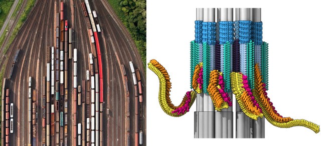
Cilia perform numerous functions for the cell: they help cells swim, move fluid, and send messages to each other. The assembly and function of cilia relies on large trains of proteins that carry important cargos out to the ciliary tip (anterograde direction) and back to the base (retrograde direction). The research team has now succeeded in examining the ciliary base in its natural environment, revealing its native 3D structure for the first time. Here, they discovered a busy transport hub, with trains being assembled and loaded in preparation for their journey into the cilia.
Loading station for cilia transport
Cilia are firmly anchored to the cell at their base. “Ciliary trains are assembled here with a very similar structure to the one that we previously showed for anterograde trains inside the cilium.” explains Gaia Pigino, Associate Head of the Structural Biology Centre at Human Technopole. “This suggests that train assembly is necessary of recruitment and loading of cargo” continues Nikolai Klena, shared first author of the study and currently a postdoc ad Human Technopole. At the ciliary base the forming trains are readily placed on the microtubular rails. There are a total of nine different rails inside cilia, called microtubule doublets. The trains are powered by specialized molecular motors and transport proteins such as signaling molecules and building materials to the tip of the cilia. At their destination station, the train is unloaded and disassembled.
The team examined the composition of the assembling trains in detail, revealing the order with which the train components are put together at the ciliary base. They also imaged structures at the base that serve as a selective barrier. “This regulates the entry of large trains until they are fully assembled and loaded with the cargo proteins required for the construction and maintenance of the cilia,” says Hugo van den Hoek, first authors of the study. “From fluorescence microscopy, we also know the exact timetable of the trains. Trains leave the start station within nine seconds, and then the whole train assembly process starts again.”
Function of the transport system
Cilia are involved in a wide variety of processes in the body. For example, they ensure that we can see, that substances are removed from the lungs, that fluid moves in the brain, and that we perceive smells and sound. They are also essential for our development and the correct arrangement of our organs. If their function is disturbed, a wide variety of diseases can result, including heart, kidney, and lung diseases, blindness or infertility. Even the smallest mutations in individual components can paralyse the traffic inside cilia. The trains are made of large complexes of numerous proteins that function as adaptors between the molecular motors and their various cargos. “In the future, the investigation of how the individual protein components of the transport machinery organize into train structures will be fundamental to understand train mechanisms and the molecular basis of ciliopathies that are associated with train malfunctions.” says Gaia Pigino.
Organelles in 3D: the techniques
The researchers resolved the structure and composition of the ciliary base with the help of complementary imaging methods.
Cryo-electron microscopy (cryo-EM) is an imaging technique that allows the analysis of biological samples (cells and molecules) at sub-nanometer resolution.
Cryo-Focused Ion Beam-Scanning Electron Microscopy (cryo-FIB-SEM) and cryo-electron tomography (cryo-ET) are cryo-EM technologies for sample preparation and imaging that allow researchers to combine 2D images of vitrified samples taken at different angles to reconstruct the 3D image (or tomogram) of intracellular organelles and protein complexes in a near-native state in their cellular environment.
Ultrastructure expansion microscopy (U-ExM) is an advanced microscopy technique developed by the Hamel’s and Guichard’s laboratories whereby it is possible to obtain super-resolution images of biological samples by using conventional optical microscopes. This technique preserves sample ultrastructure after expansion.
Publication
Hugo van den Hoek, Nikolai Klena, Mareike A. Jordan, Gonzalo Alvarez Viar, Ricardo D. Righetto, Miroslava Schaffer, Philipp S. Erdmann, William Wan, Stefan Geimer, Jürgen M. Plitzko, Wolfgang Baumeister, Gaia Pigino, Virginie Hamel, Paul Guichard, Benjamin D. Engel: In situ architecture of the ciliary base reveals the stepwise assembly of IFT trains. Science, published online.
Share:
You could also like:
-
HT National Facilities: One Year On, New Call Open
On 10 June, Human Technopole marks the one-year anniversary of the official opening of its National Facilities, a key milestone in our mission to support excellence in life sciences across Italy. Celebrating this important date, we are pleased to announce that the new Call for Access to the National Facilities is now open. Researchers can submit their proposals until 30 September 2025.
-
10 Fully Funded PhD Scholarships in Systems Medicine through SEMM
Human Technopole is offering up to 10 fully funded PhD scholarships through the SEMM PhD Program in Systems Medicine. These scholarships are open to talented and motivated graduates—both from Italy and abroad – interested in pursuing doctoral research.
-

Human Technopole Commitment to Gender Equality
Human Technopole is proud to announce the launch of its new Gender Equality Plan (GEP) for 2025-2027, a strategic initiative designed to further integrate gender equality across all aspects of the Institute, building upon the successful outcomes of the previous GEP (2022-2024). HT has also achieved the UNI/PdR 125:2022 certification, which recognizes organizations that promote gender equality and foster inclusive workplaces.
-
2 fully funded PhD fellowships at HT through SISSA
Human Technopole is offering 2 fully funded 4-year PhD fellowships to young scientists from the national and international community who wish to undertake a doctoral degree on a project focused on Computational Biology.
-

Childhood Cancer: Free DNA in Blood Reveals Therapy Resistance
An international study coordinated by Milan’s Human Technopole, the Institute of Cancer Research (ICR) in London, and the Royal Marsden Hospital in London has shown that certain DNA fragments found in the blood of paediatric cancer patients can be used as “biomarkers” to obtain information on the characteristics of the disease and its ability to resist therapies. Analysing these fragments could represent an effective alternative to tumour tissue biopsy, a practice that is particularly difficult in children.