Ultrastructure Expansion Microscopy details Chlamy native organisation
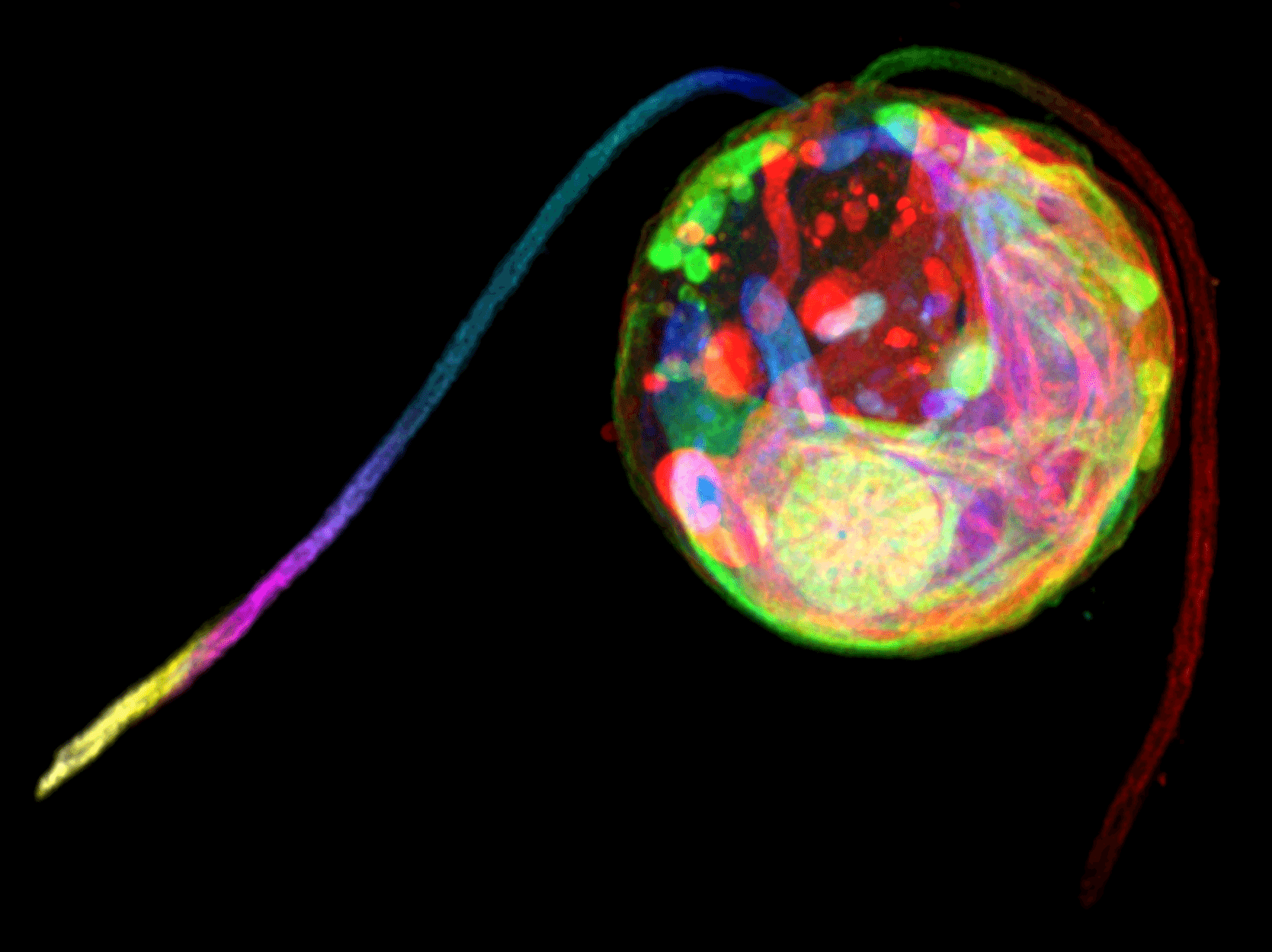
The Pigino Group developed a detailed protocol to obtain super-resolution images of the green alga Chlamydomonas reinhardtii by using conventional optical light microscopes.
The advent of super-resolution imaging techniques coupled with fluorescence microscopy allowed the visualisation of a vast array of biological samples with a resolution higher than that imposed by the light diffraction limit. However, super-resolution microscopes are usually expensive instruments with complicated setups and require experienced personnel for sample preparation and image acquisition. Expansion Microscopy (ExM) has recently been developed to overcome these limitations and uses swellable hydrogels to obtain up to fourfold isotropic expansion of the sample before imaging with a confocal microscope.
Gaia Pigino, Associate Head of the Structural Biology Research Centre at Human Technopole, Nikolai Klena and Giovanni Maltinti, members of the Pigino Group, in collaboration with Paul Guichard and Virginie Hamel at the University of Geneve, developed an ExM-based protocol to visualise the three-dimensional (3D) ultrastructural organisation of Chlamydomonas reinhardtii (Ultrastructure Expansion Microscopy, U-ExM). The protocol is now published in the open-access journal Bio-protocol and made available to the research community.
Chlamydomonas is a single-cell model organism widely used to investigate the molecular mechanisms of cilia and flagella motility. In this protocol, the researchers compare different fixation and staining procedures and provide instructions on how to embed the sample in a hydrogel and acquire images with a confocal microscope as well as how to analyse them.
The Pigino Group has already used this protocol to study the assembly of intraflagellar transport trains (IFT) at the Chlamydomonas ciliary base1, thus showing that this new procedure will be instrumental for researchers to perform super-resolution analysis of target proteins in a native 3D environment without the need for expensive super-resolution microscopes and specific training.
1 van den Hoek et al. (2022) In situ architecture of the ciliary base reveals the stepwise assembly of intraflagellar transport trains. Science
Share:
You could also like:
-
HT National Facilities: One Year On, New Call Open
On 10 June, Human Technopole marks the one-year anniversary of the official opening of its National Facilities, a key milestone in our mission to support excellence in life sciences across Italy. Celebrating this important date, we are pleased to announce that the new Call for Access to the National Facilities is now open. Researchers can submit their proposals until 30 September 2025.
-
10 Fully Funded PhD Scholarships in Systems Medicine through SEMM
Human Technopole is offering up to 10 fully funded PhD scholarships through the SEMM PhD Program in Systems Medicine. These scholarships are open to talented and motivated graduates—both from Italy and abroad – interested in pursuing doctoral research.
-

Human Technopole Commitment to Gender Equality
Human Technopole is proud to announce the launch of its new Gender Equality Plan (GEP) for 2025-2027, a strategic initiative designed to further integrate gender equality across all aspects of the Institute, building upon the successful outcomes of the previous GEP (2022-2024). HT has also achieved the UNI/PdR 125:2022 certification, which recognizes organizations that promote gender equality and foster inclusive workplaces.
-
2 fully funded PhD fellowships at HT through SISSA
Human Technopole is offering 2 fully funded 4-year PhD fellowships to young scientists from the national and international community who wish to undertake a doctoral degree on a project focused on Computational Biology.
-

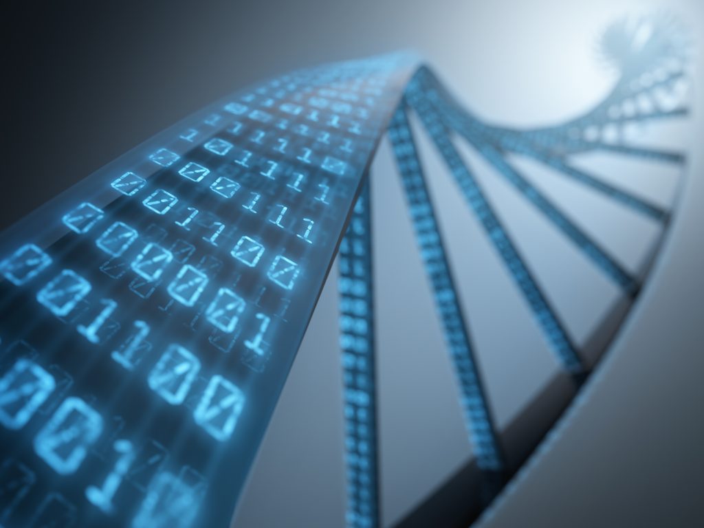
Childhood Cancer: Free DNA in Blood Reveals Therapy Resistance
An international study coordinated by Milan’s Human Technopole, the Institute of Cancer Research (ICR) in London, and the Royal Marsden Hospital in London has shown that certain DNA fragments found in the blood of paediatric cancer patients can be used as “biomarkers” to obtain information on the characteristics of the disease and its ability to resist therapies. Analysing these fragments could represent an effective alternative to tumour tissue biopsy, a practice that is particularly difficult in children.