
Pigino Group
Cells need to be able to sense different types of signals, such as chemical and mechanical signals, from the extracellular environment to properly function. Most eukaryotic cells perform these functions through a specialized hair-like organelle, the cilium, that extends from the cell body as a sort of antenna. The signaling and sensory functions of cilia are fundamental already during the early stages of embryo development, when cilia coordinate the establishment of the internal left/right asymmetry typical of the vertebrate body. Later, cilia continue to be required for the correct development and function of specific tissues and organs, such as brain, heart, kidney, liver, and pancreas. Sensory cilia eventually allow us to sense the environment that surrounds us, for instance we see through the connecting cilium of photoreceptors in our retina, we smell through the sensory cilia at the tip of our olfactory neurons, and we hear thanks to the kinocilium of our sensory hair cells. Motile cilia, which themselves have sensory functions, also work as propeller-like extensions that allow us to breath, because they keep our lungs clean, to reproduce, because they propel sperm cells, and even to properly reason, because they contribute to the flow of cerebrospinal fluid in our brain ventricles. Not surprisingly, defects in the assembly and function of these tiny organelles result in devastating pathologies, which are collectively known as ciliopathies. Thus, proper function of cilia is fundamental for human health.
The Pigino Lab investigates the biology and the 3D molecular structure of ciliary components in their native cellular context and in isolation, to understand how they orchestrate cilia-specific functions. Our work positions itself right at the interface between structural biology and molecular cell biology. Hence, we combine the latest tools and methodologies from both fields, from cryo-electron tomography, over correlative light and fluorescence microscopy (CLEM), to in vitro reconstituted dynamic systems, genetics, biochemistry, image analysis methods, all the way to more classical cell biology.
Our ultimate goal is to understand the underlying molecular mechanisms of ciliary functions and dysfunctions, so that possible therapeutic strategies for ciliopathies can be developed.

ERC funded project (ERC-2018-COG N#819826 CiliaTubulinCode)
Self-organization of the cilium: the role of the tubulin code
Our current knowledge of the basic principles which lead to self-organization of cellular organelles is quite limited. Hence, our project aims at understanding the role of the tubulin code for self-organization of complex microtubule based cellular structures, with the focus on the structure of the cilium.
Group members
-
 Gaia Pigino
Associate Head of Structural Biology Research Centre -
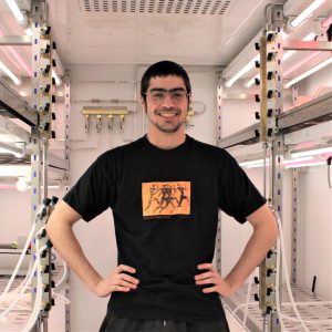 Gonzalo Alvarez Viar
Postdoc -
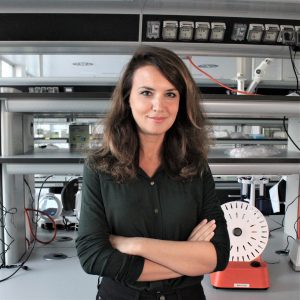 Paola Capasso
Senior Technician -
 Rubina Dad
Postdoc -
 Helen Elizabeth Foster
Postdoc -
 Malina Karen Iwanski
Postdoc -
 Nikolai Klena
Postdoc -
 Samuel Edward Lacey
Postdoc -
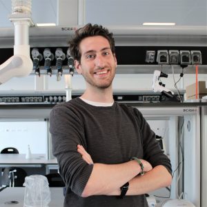 Giovanni Maltinti
PhD Student -
 Adrian Pascal Nievergelt
Scientific Visitor
Publications
-
10/2015 - Journal of Structural Biology
Three-dimensional mass density mapping of cellular ultrastructure by ptychographic X-ray nanotomography
We demonstrate absolute quantitative mass density mapping in three dimensions of frozen-hydrated biological matter with an isotropic resolution of 180 nm. As model for a biological system we use Chlamydomonas cells in buffer solution confined in a microcapillary. We use ptychographic X-ray computed tomography to image the entire specimen, including the 18 μm-diameter capillary, thereby […]
-
05/2015 - Optics Express
Artifact characterization and reduction in scanning X-ray Zernike phase contrast microscopy
Zernike phase contrast microscopy is a well-established method for imaging specimens with low absorption contrast. It has been successfully implemented in full-field microscopy using visible light and X-rays. In microscopy Cowley’s reciprocity principle connects scanning and full-field imaging. Even though the reciprocity in Zernike phase contrast has been discussed by several authors over the past […]