
Pigino Group
Cells need to be able to sense different types of signals, such as chemical and mechanical signals, from the extracellular environment to properly function. Most eukaryotic cells perform these functions through a specialized hair-like organelle, the cilium, that extends from the cell body as a sort of antenna. The signaling and sensory functions of cilia are fundamental already during the early stages of embryo development, when cilia coordinate the establishment of the internal left/right asymmetry typical of the vertebrate body. Later, cilia continue to be required for the correct development and function of specific tissues and organs, such as brain, heart, kidney, liver, and pancreas. Sensory cilia eventually allow us to sense the environment that surrounds us, for instance we see through the connecting cilium of photoreceptors in our retina, we smell through the sensory cilia at the tip of our olfactory neurons, and we hear thanks to the kinocilium of our sensory hair cells. Motile cilia, which themselves have sensory functions, also work as propeller-like extensions that allow us to breath, because they keep our lungs clean, to reproduce, because they propel sperm cells, and even to properly reason, because they contribute to the flow of cerebrospinal fluid in our brain ventricles. Not surprisingly, defects in the assembly and function of these tiny organelles result in devastating pathologies, which are collectively known as ciliopathies. Thus, proper function of cilia is fundamental for human health.
The Pigino Lab investigates the biology and the 3D molecular structure of ciliary components in their native cellular context and in isolation, to understand how they orchestrate cilia-specific functions. Our work positions itself right at the interface between structural biology and molecular cell biology. Hence, we combine the latest tools and methodologies from both fields, from cryo-electron tomography, over correlative light and fluorescence microscopy (CLEM), to in vitro reconstituted dynamic systems, genetics, biochemistry, image analysis methods, all the way to more classical cell biology.
Our ultimate goal is to understand the underlying molecular mechanisms of ciliary functions and dysfunctions, so that possible therapeutic strategies for ciliopathies can be developed.

ERC funded project (ERC-2018-COG N#819826 CiliaTubulinCode)
Self-organization of the cilium: the role of the tubulin code
Our current knowledge of the basic principles which lead to self-organization of cellular organelles is quite limited. Hence, our project aims at understanding the role of the tubulin code for self-organization of complex microtubule based cellular structures, with the focus on the structure of the cilium.
Group members
-
 Gaia Pigino
Associate Head of Structural Biology Research Centre -
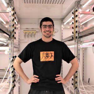 Gonzalo Alvarez Viar
Postdoc -
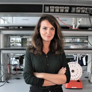 Paola Capasso
Senior Technician -
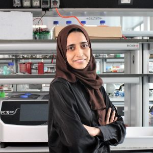 Rubina Dad
Postdoc -
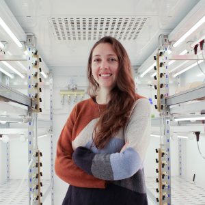 Helen Elizabeth Foster
Postdoc -
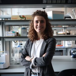 Malina Karen Iwanski
Postdoc -
 Nikolai Klena
Postdoc -
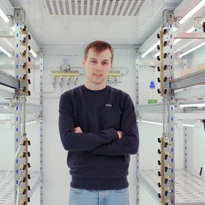 Samuel Edward Lacey
Postdoc -
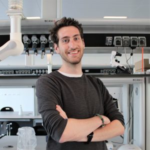 Giovanni Maltinti
PhD Student -
 Adrian Pascal Nievergelt
Scientific Visitor
Publications
-
10/2017 - Molecular Biology of the Cell
Microtubule dynamics: 50 years after the discovery of tubulin and still going strong
The Minisymposium “Microtubule Dynamics” featured speakers ranging from a founding member of the microtubule field to graduate students embarking on their scientific journeys. The session spanned a broad range of topics, from fundamental questions about the structure and dynamics of microtubules, to the behavior of molecular motors in reconstituted systems, to the therapeutic potential of […]
-
07/2017 - Nature Structural & Molecular Biology
Switching dynein motors on and off
Cytoplasmic dyneins transport cellular components from the periphery toward the center of the cell. By moving cargoes along microtubules, dyneins ensure proper cell division, regulate exchange of materials between organelles, and contribute to the internal organization of eukaryotic cells. Two recent studies show that, upon dimerization, cytoplasmic dyneins intrinsically adopt an autoinhibited configuration that can […]
-
04/2017 - Methods in Cell Biology
Millisecond time resolution correlative light and electron microscopy for dynamic cellular processes
Molecular motors propel cellular components at velocities up to microns per second with nanometer precision. Imaging techniques combining high temporal and spatial resolution are therefore indispensable to understand the cellular mechanics at the molecular level. For example, intraflagellar transport (IFT) trains constantly shuttle ciliary components between the base and tip of the eukaryotic cilium. 3-D […]
-
05/2016 - Science
Microtubule doublets are double-track railways for intraflagellar transport trains
The cilium is a large macromolecular machine that is vital for motility, signaling, and sensing in most eukaryotic cells. Its conserved core structure, the axoneme, contains nine microtubule doublets, each comprising a full A-microtubule and an incomplete B-microtubule. However, thus far, the function of this doublet geometry has not been understood. We developed a time-resolved […]
-
10/2015 - American Journal of Respiratory Cell and Molecular Biology
DNAH11 Localization in the Proximal Region of Respiratory Cilia Defines Distinct Outer Dynein Arm Complexes
Primary ciliary dyskinesia (PCD) is a recessively inherited disease that leads to chronic respiratory disorders owing to impaired mucociliary clearance. Conventional transmission electron microscopy (TEM) is a diagnostic standard to identify ultrastructural defects in respiratory cilia but is not useful in approximately 30% of PCD cases, which have normal ciliary ultrastructure. DNAH11 mutations are a common cause […]